Expertise
Clinical excellence in women’s health
Mentorship
Teaching that transforms
Leadership
Shaping the future of care for women
Innovation
Formulating solutions that support women


Start with the topics people search for most
Evidence-based articles and clinical context on common women’s health concerns.

Develop Expert Knowledge and Practical Skills in Women’s Health
The Institute of Women’s Health and Integrative Medicine is an educational and research organization whose mission is to provide advanced training to primary health care practitioners and to conduct and support clinical research in women’s health and natural therapies and integrative medicine.
")
Residency opportunities for Naturopathic Physicians
The priority of the Naturopathic Education and Residency Consortium (NERC) is providing additional naturopathic residencies that will help to deliver highly competent Naturopathic Physicians who can practice in a diverse array of situations, and be successful.
Health Support for Women by a Woman
Vitanica specializes in natural health products & information for women. Our formulas are clinically developed by Dr. Tori Hudson and utilized by physicians. Consistent quality is one of our commitments to you.
Latest Articles

| January 28, 2026
Polycystic ovary syndrome (PCOS) affects 5%-15% of reproductive age women. [...]
| January 14, 2026
Pelvic exams often include the use of a vaginal speculum. [...]
| December 22, 2025
Yesterday, December 21, was the winter solstice and the first [...]
| December 16, 2025
Please join me for my conversation I had with a [...]
| November 25, 2025
Danielle Melvin N.D. guest author The FDA has announced it [...]
| November 17, 2025
The American Heart Association (AHA) and the American College of [...]

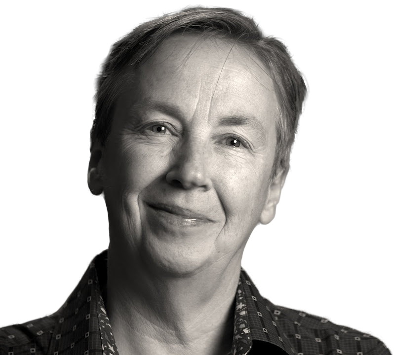
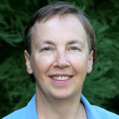
Meet Dr. Tori Hudson
For more than 40 years, Dr. Tori Hudson, Naturopathic Physician, has helped women move from dissatisfaction, confusion and conflicting health advice to scientific, evidence-backed guidance they can trust. A trailblazer in integrative women’s health, she blends rigorous scientific research, traditional naturopathic wisdom, and decades of clinical experience to offer practical, individualized care, and to mentor the next generation of clinicians who will carry this work forward.
Subscribe to the Blog
Occasional updates with new articles, education, and media. No hype.
